A brain imaging study reveals how the brain decodes the social significance of facial ornaments
Published on the CNRS biology website: https://www.insb.cnrs.fr/fr/cnrsinfo/quand-le-cerveau-decode-la-signification-sociale-des-ornements-du-visage
Since prehistoric times, we have decorated our faces with ornaments and paintings to communicate our social role. An interdisciplinary group of researchers from the University of Bordeaux and the CNRS have used functional magnetic resonance imaging (fMRI) to map the brain regions involved in attributing social status to faces decorated with ornaments and paintings. This research sheds light on the brain mechanisms underlying the interpretation of social markers on the human face and provides valuable insights into the cultural evolution of our species. It has just been published in Brain Structure and Function.
For at least 150,000 years, humans have been culturally modifying their bodies by wearing ornaments and using pigments. These practices have been powerful means of communication, expressing identity, group membership, and social status. To identify the brain processes associated with attributing social status to facial ornaments, an interdisciplinary team including neuroscientists from the Neurofunctional Imaging Group (Institute of Neurodegenerative Diseases, University of Bordeaux – CEA – CNRS) and archaeologists from the PACEA laboratory (University of Bordeaux – CNRS – Ministry of Culture) explored the mechanisms at work in the human brain, using fMRI.
The study results reveal that several brain regions are involved in the encoding, evaluating, and categorizing of social status based on facial ornamentation. The fusiform gyrus, known for its role in face perception, is crucial in this activity, enabling individuals to recognize and process faces as vectors of social information. The orbitofrontal cortex, associated with social evaluation and decision-making, is also activated when attributing social status.
The salience network, responsible for detecting and integrating relevant information, also showed significant activity during this task.
Another notable result is the activation of the hippocampus and parahippocampus, regions associated with memory and associative abilities. These regions probably facilitate recalling past experiences and retrieving relevant information needed to make social judgments based on face ornamentation. The inferior frontal gyrus, a region involved in language processing, also showed activity while interpreting facial ornaments as vectors of information about individuals’ social status. This result suggests proximity between processes linked to language comprehension and those applied to the symbolic interpretation of facial ornamentation.
An analysis of functional connectivity in the resting state revealed complex interactions and coordination between the fusiform gyrus, orbitofrontal cortex, salience network, hippocampus, parahippocampus, and inferior frontal gyrus. These brain networks collectively contribute to interpreting and attributing social status based on facial ornamentation.
The results of this study represent a significant advance in our understanding of the cerebral underpinnings of social status attribution and the symbolic interpretation of social markers on the human face. The researchers propose that these connections must already have been partly in place in Homo populations using red ochre 300,000 years ago and must have reached a degree of integration close to our own 140,000 years ago in Homo sapiens, who combined the use of ochre for body painting with the use of shell ornaments.
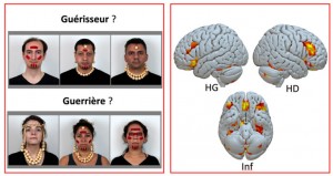
Left: Based on facial ornamentation, participants had to choose the person who best matched the proposed status from three possibilities. Right: Lateral and inferior views of brain activations triggered by the attribution of social status. (LH: left hemisphere, RH: right hemisphere, Inf: inferior view).
Reference
Salagnon M., d’Errico F., Rigaud S. Mellet E. 2024. Assigning a social status from face adornments: an fMRI study.
Brain Structure and Function
https://link.springer.com/article/10.1007/s00429-024-02786-4
Contact
Emmanuel Mellet – emmanuel.mellet@u-bordeaux.fr / +33 5 33 51 48 43
IMN UMR 5293 – GIN E5
CEA – CNRS – Université de Bordeaux
PAC Carreire – Case 28
146 rue Léo Saignat CS 61292
33076 Bordeaux Cedex
Francesco d’Errico – francesco.derrico@u-bordeaux.fr / +33 5 40 00 26 28
PACEA UMR 5199
CNRS – Université de Bordeaux
Bâtiment B2
Allée Geoffroy Saint Hilaire
CS 50023 33615 PESSAC CEDEX


